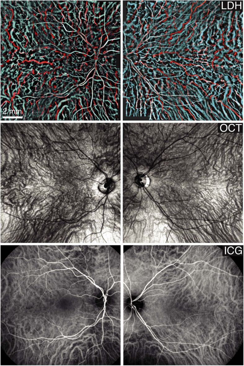
Fig. 5.
Imaging the choroid with LDH, OCT (Plex Elite 9000, Zeiss), and ICG-angiography (Spectralis, Heidelberg). The LDH montages have been stitched from 5x5 images obtained by fusing the 2-6 and 10-30 kHz frequency ranges in cyan and red. The access to the low frequency range made routinely possible by SVD is critical to reveal choroidal veins.

