Fig. 13.

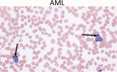
Peripheral blood showing two myeloblasts with a high ratio of nucleus to cytoplasm, finely dispersed chromatin and one or more large nucleoli (arrows). Acute myeloid leukemia represents only 20 % of childhood leukemia

Peripheral blood showing two myeloblasts with a high ratio of nucleus to cytoplasm, finely dispersed chromatin and one or more large nucleoli (arrows). Acute myeloid leukemia represents only 20 % of childhood leukemia