Fig. 9.

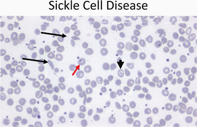
Peripheral smear (40 ×) from a patient with sickle cell disease showing sickle cells (black arrows), target cells (arrowhead), and a Howell–Joley body (red arrow)

Peripheral smear (40 ×) from a patient with sickle cell disease showing sickle cells (black arrows), target cells (arrowhead), and a Howell–Joley body (red arrow)