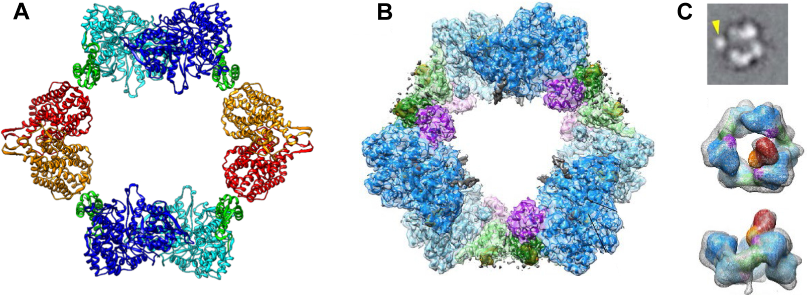
Figure 6.

Structures of dATP inhibited states of class Ia RNRs. A X-ray structure of dATP inhibited E. coli class Ia RNR (29) is an α4β4 ring structure with a hole in the middle, composed of alternating α2 (light and dark blue with the cone domains in green) and β2 (orange and red) subunits. Note the importance of the cone domain in the α/β interaction. B Cryo-EM structure of dATP inhibited human class Ia RNR (27) is a hexameric α6 ring with a hole in the middle. A subunits are in light blue and dark blue with cone domain in light and dark green and a three-helix insertion in purple (residues 638–681). Note the importance of the cone domain in the α/α interactions. C) Cryo-EM structure of α/β (1:1)clofarabine triphosphate (ClFTP) inhibited human class Ia RNR (27). Top panel is a representative cryo -EM 2D class average image generated from α, β and clofarabine triphosphate (ClFTP) that shows β (arrow) interacting with α. The middle and bottom panels are two views of the 3D reconstruction of the same data set. The bottom is rotated 90° from the middle image. Only a fraction of the α6 rings in these images have a single and variably positioned β.
