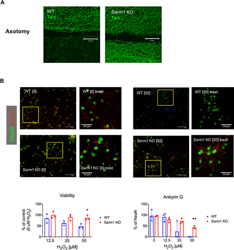
Fig 1. Delayed axonal degeneration phenotype of Sarm1 knockout (KO) neurons in culture.

(A) Cultured neurons from wild type (WT) and Sarm1 KO mice were subjected to axotomy, then labeled for Tau 72 hours later. Confocal microscopy of Tau immunocytochemistry shows loss of Tau in distal axonal segments in WT, but not Sarm1 KO, neurons at 72 h following axotomy. Representative images from 3 independent experiments. Scale bar, 50 μm. (B) Cultured neurons from WT and Sarm1 KO mice were subjected to oxidative stress by 30 min exposure to 0, 12.5, 25 or 50 μM hydrogen peroxide (H2O2) then assayed 24 h later. Images show ankyrin G (red) and NeuN (green), detected by immunocytochemistry in cultured neurons from WT and Sarm1 KO mice. Scale bar, 100 μm and inset scale bar 50 μm. Bar graph (left) shows mean neuronal viability +/- standard deviation at indicated H2O2 concentrations, assessed by dye exclusion and normalized to control (0 μM H2O2). * denotes p = 0.011, t-test with Holm-Sidak method for multiple comparisons. Bar graph (right) shows mean ankyrin G expression (% of NeuN) +/- standard deviation. ** denotes p < 0.001, t-test with Holm-Sidak method for multiple comparisons.
