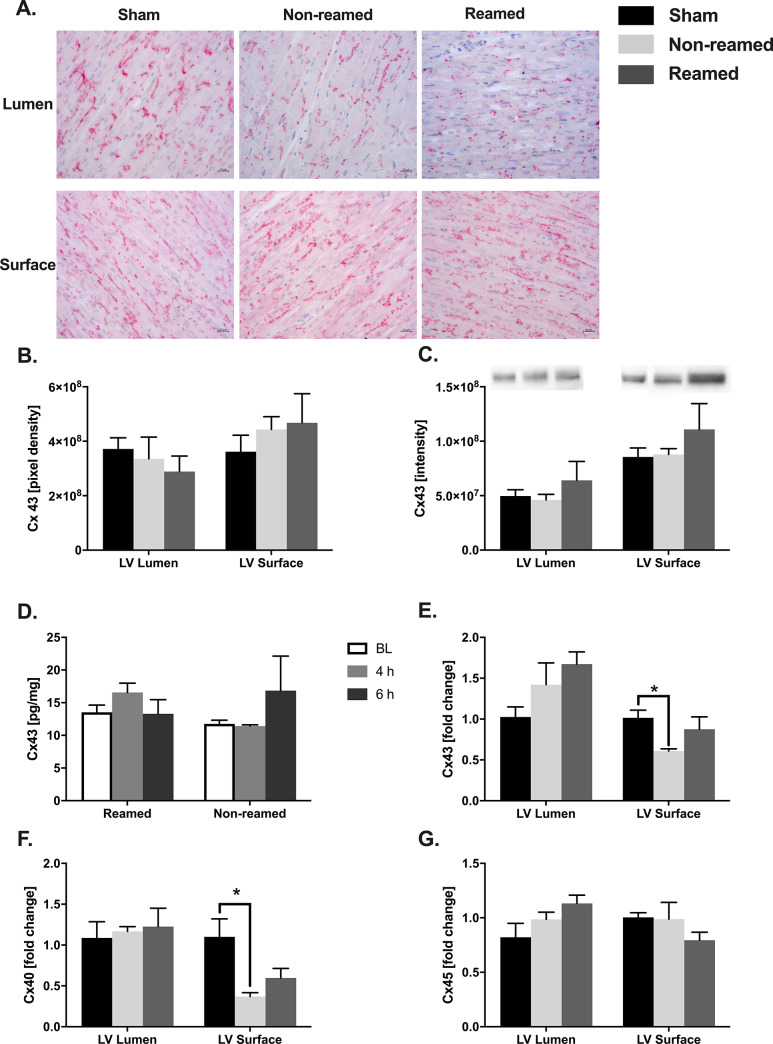
Fig 1. Alteration of connexin 43 distribution after multiple trauma in pigs.
A) Representative images of connexin 43 (Cx43) in the superficial and luminal layer of the left ventricle (LV) after multiple trauma and either nailing (non-reamed) or reaming. B) Amount of Connexin 43 measured in stained sections of the cardiac tissue after multiple trauma in pigs. Control/sham animals were presented as black bars, pigs after multiple trauma and nailing (non-reamed) were presented as light grey and pigs after multiple trauma and reaming (reamed) were presented as dark grey bars. C) Western blot analysis of Cx43 protein expression, as well as representative western blot bands. D) Systemic levels of connexin 43 in pg/mg measured in serum taken at baseline (BL), 4h and 6h after trauma. Baseline amounts are represented as white bars, 4 h after trauma as medium grey bars and 6 h after trauma as dark grey bars. E) Cx43 mRNA-expression in the LV presented as fold change. F) Connexin 40 (Cx40) mRNA expression detected in tissue homogenates of the left ventricle 6h after multiple trauma. G) Connexin 45 (Cx45) mRNA expression presented as fold change compared to sham. Results are significant (*) p<0.05. For statistical analysis one-way ANOVA was used. Graphical presentation as mean ± SEM. For each experiment n = 5.

