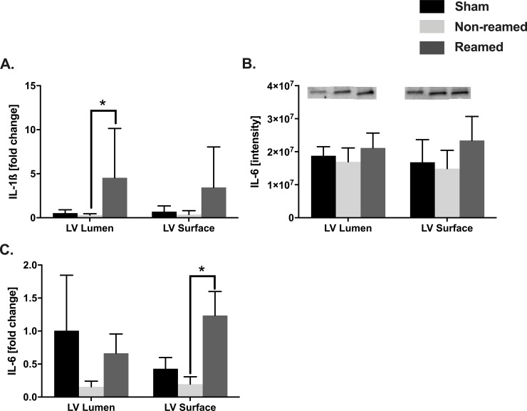
Fig 3. Cardiac inflammation.
Control/sham animals were presented in black bars, pigs after multiple trauma and nailing (non-reamed) were presented in light grey and pigs after multiple trauma and reaming (reamed) were presented as dark grey bars. A) mRNA expression of interleukin 1-ß (IL-1ß) of the luminal and superficial layer of the left ventricle, detected by RT-qPCR presented as fold change. B) Interleukin-6 (IL-6) protein expression and representative western blot bands. C) mRNA expression of IL-6 in the left ventricle. n = 5 for each experiment. Results are significant (*) p<0.05. For statistical analysis one-way ANOVA was used. Graphical presentation as mean ± SEM.

