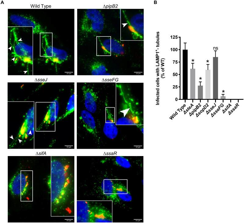
Fig 1. LAMP1+-tubule extension from single deletion mutants.
(A) Comparison of frequency of LAMP1+-tubule formation of WT and isogenic single-effector deletion mutants in HeLa cells after 8 hours of infection. Cells were immunostained for Salmonella (red) and LAMP1 (green), and the nucleus was stained with DAPI (blue). Representative images of select strains are shown. The white boxes indicate zoomed-in region in inset. Arrowheads indicate LAMP1+-tubules. Scale Bar = 10 μm. (B) Quantification of LAMP1+-tubule frequency in HeLa cells infected with the single deletion mutants for 8 hours. The average frequency of infected cells with LAMP1+-tubules relative to wild type infected cells ± standard error of the mean for three separate experiments is shown (n = 3). At least 100 infected cells per strain were blindly analyzed in each experiment. An asterisk indicates a significant difference between the indicated mutant strain LAMP1+-tubule frequency and the corresponding WT LAMP1+-tubule frequency (p < 0.02) as determined by Kruskal-Wallis one-way ANOVA with Dunn’s multiple comparison post-test.

