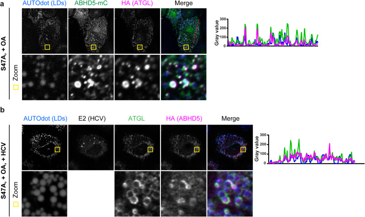
Fig 3. ABHD5 and ATGL colocalize at the lipid droplet surface.
ABHD5 and ATGL S47A were expressed by lentiviral transduction and detected by indirect immunofluorescence using antibodies against the respective epitope tag or by their auto-fluorescence in case of the ABHD5-mCitrine fusion protein. Colocalization between the proteins or with the lipid droplet marker AUTOdot was tested in naive (a) or HCV-infected (b) Lunet N hCD81 cells after oleic acid (OA) induction. (a) Subcellular localization of HA-tagged ATGL S47A relative to ABHD5-mCitrine (mC) and lipid droplets. (b) Subcellular localization of HA-tagged ABHD5 relative to ATGL S47A and lipid droplets, in HCV-infected cells. HCV infection was verified by immunostaining against HCV E2 glycoprotein. In (a) and (b) the plots on the right side indicate the intensity profiles in the different channels along the red dotted line depicted in the merge picture.

