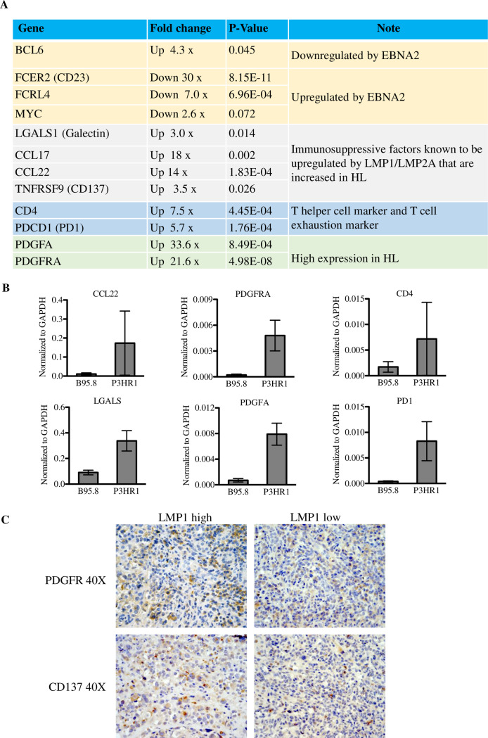
Fig 10. P3HR1 infected lymphomas express cellular genes upregulated in RS cells of Hodgkin lymphomas.
RNA was isolated from tumors infected with B95.8 or P3HR1 virus-induced lymphomas, and RNA-seq performed. Mouse cell transcripts were removed from further analysis, and the levels of human genes in each tumor type was compared as described in the methods. A. The relative levels of cellular gene transcripts in P3HR1-infected tumors, versus B95.8-infected tumors, are shown for known EBNA2 target genes (yellow shading), immunosuppressive factors which are highly expressed in RS cells and are known LMP1 targets (gray shading), markers for helper T cells and T cell exhaustion (blue shading), and markers for enhanced PDGF signaling (which is often increased in HL tumors)(green shading). The fold-change in cellular gene expression in P3HR1-infected tumors versus B95.8-infected tumors is indicated, as well as the p-value for each difference. B. qPCR was performed using cDNA isolated from two different P3HR1 virus-induced lymphomas versus two different B95.8 virus-infected lymphomas, using human specific primers to amplify genes. Results were normalized to the level of GAPDH transcript. Standard error is shown. C. IHC analysis using antibodies against PDGFRA or CD137 was performed in areas of tumors with low or high level LMP1 expression. Both P3HR1 tumor sections shown are derived from mouse #2 (S1 Table).

