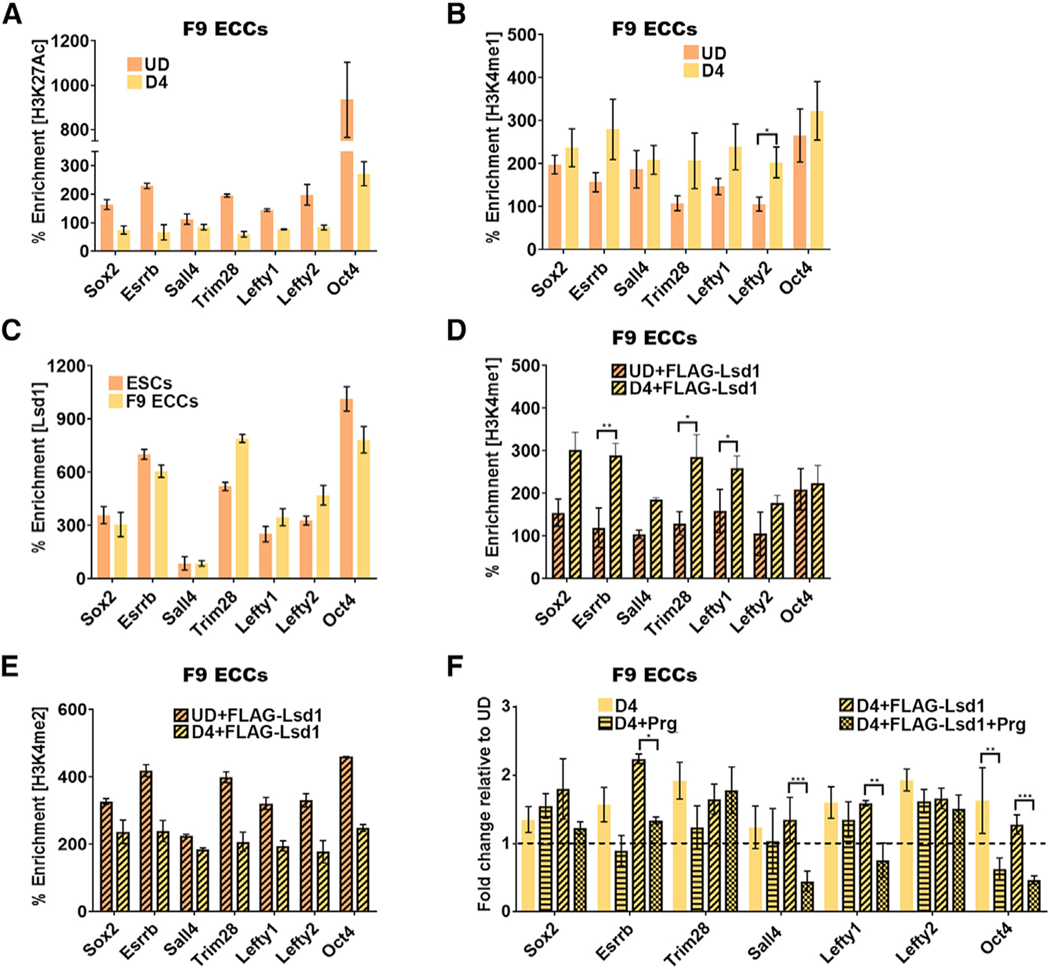
Figure 3. A “Primed” Chromatin State Is Established at Pluripotency Gene Enhancers in Embryonal Carcinoma Cells.

Chromatin immunoprecipitation (ChlP)-qPCR assays.
(A and B) Histone modifications at PpGe (A) H3K27Ac and (B) H3K4me1 in F9 ECCs pre- and D4 post-differentiation. Whereas deacetylation of PpGe is observed as a decrease in the H3K27Ac signal, histone H3K4me1 is retained post-differentiation.
(C) Lsd1 occupancy in undifferentiated ESCs and F9 ECCs.
(D and E) Enrichment of (D) H3K4me1 and (E) H3K4me2 in F9 ECCs expressing recombinant FLAG-Lsd1 compared to undifferentiated transfected cells. The ECCs were differentiated 24 h post-transfection with Lsd1-expressing plasmid. Whereas there was an increase in H3K4me1 at some PpGe (D), there was a concomitant decrease in H3K4me2 enrichment (E).
(F) Fold change in enrichment of H3K4me1 at PpGe in pargyline treated and untreated, WT, and FLAG-Lsd1 overexpressing F9 ECCs at D4 post-differentiation. Fold change is represented as relative to enrichment in the undifferentiated state (dotted line).
% Enrichment = fold enrichment over input x 100. p values were derived from Student’s t test: *p < 0.05; **p < 0.01; ***p < 0.005. UD, undifferentiated; D4, days post-induction of differentiation; D4+FLAG-Lsd1, F9 ECCs overexpressing FLAG- Lsd1 and differentiated for 4 days; Prg, pargyline; pluripotency gene enhancers. All experiments are an average of atleast two biological replicates and error is shown as SEM.
