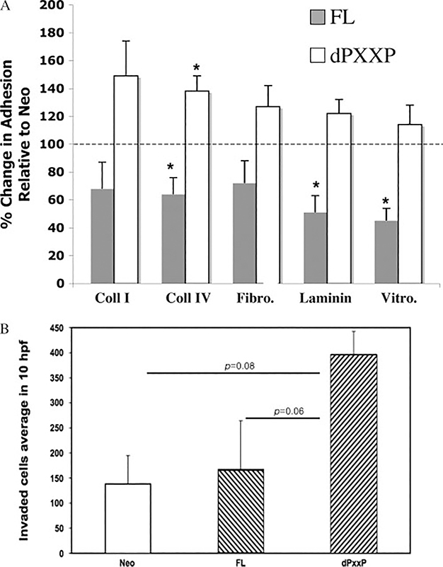
Figure 1.
dPXXP-overexpressing cells are more adhesive and more invasive in MDA435 cells. (A) Vector control, FL, and dPXXP cells were plated onto extracellular matrix-coated plates, allowed to adhere for 3 h, and then stained and assessed. Results represent the % change relative to Neo (100%, represented by a horizontal line) of at least three independent experiments (± SEM). ∗p < 0.05. (B) Net invasion is affected by overexpression of dPXXP. Corrected invasion was highest in dPXXP cells (p= 0.008). A trend towards greater invasion of dPXXP cells over FL is seen (p= 0.06)

