Figure 3.
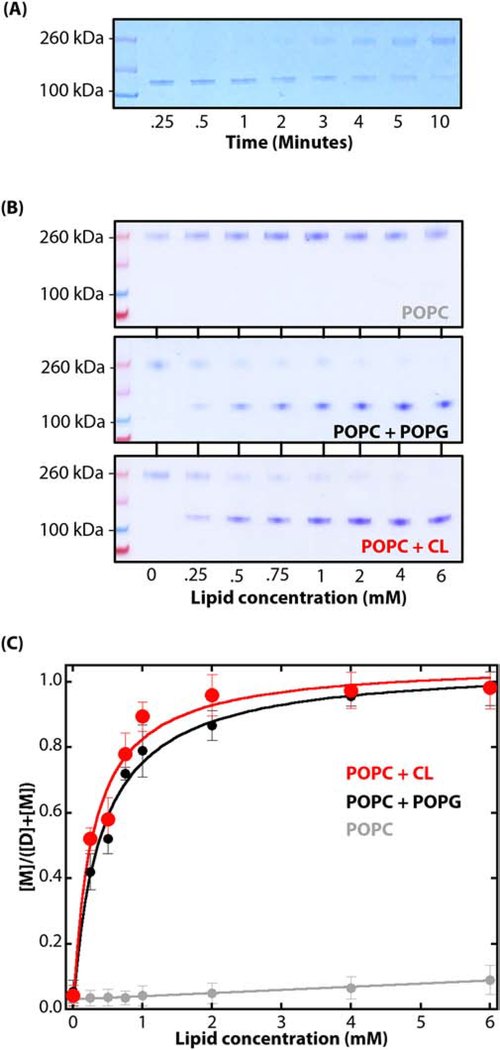
Dimeric SecA dissociates into monomers upon binding to LUV. (A) In the presence of LUV, there is a progressive shift from monomers to dimers with crosslinking time. After binding to 6 mM LUV (POPC:POPG:CL=0.7:0.2:0.1) for 30 minutes at 37°C, GA was introduced (0.15%) and allowed to crosslink for the periods shown before quenching the reaction. We conclude that as the cross-linking time increases, there is a shift from membrane-bound monomers to unbound dimers in solution. (B) Coomassie-blue stained SDS-PAGE denaturing gels showing the results of titrating SecA (1 μM) with lipid vesicles made of POPC (grey), POPC:POPG (0.7:0.3, black), or POPC:CL (0.7:0.3, red). In each case, partitioning was allowed for 30 minutes at 37°C before introducing GA (0.15%) for 15 seconds before stopping the reaction by the addition of an excess of 100 mM Tris-HCl pH 7.0). Note the progressive shift from dimers to monomers with increases in lipid concentration. (C) The changes in relative intensities of the bands on the gel as the lipid concentration is increased allows one to compute the fraction of SecA partitioned into the LUV at a given lipid concentration. The fraction partitioned fP is given by [M]/([M] + [D]) where [M] is given by the intensity of the monomer band and [D] is the intensity of dimer band. The color code is the same as panel B. The water-to-bilayer free energies of transfer ΔGwb for LUV formed from POPC:POPG and POPC:CL as −7.0 ± 0.3 and −7.1 ± 0.2 kcal.mol−1, respectively. These values agree well with the value of ΔGwb determined by fluorescence measurements of SecA partitioning into LUV formed from E. coli lipids (−7.4 ± 0.1 kcal mol−1) [10]

