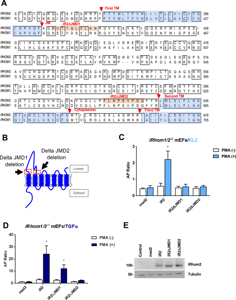
Figure 3. Characterization of the role of the iR2 extracellular JMD 1 or 2 on stimulated ADAM17-dependent shedding.
(A) Amino acid sequence alignment of partial sequences of iR1 and iR2, starting with a part of the N-terminal cytoplasmic domain, followed by the TMD1, the extracellular loop domain, and TMD2 and a part of TMD3. Identical sequences are boxed and transmembrane domains are shaded in light blue. The extracellular juxtamembrane sequences that are deleted in iR2ΔJMD1 and iR2ΔJMD2 are highlighted with orange dots. (B) Schematic indicating the position of the deleted regions in the iR2 juxtamembrane domain in iR2ΔJMD1 and iR2ΔJMD2. (C, D) Ectodomain shedding assays in iR1/2−/− mEFs after co-transfection of the iR2-dependent substrate AP-tagged Kit-ligand 2 (KL2, C) or AP-tagged TGFα (D) along with iR2, iR2ΔJMD1 and iR2ΔJMD2. Results are shown as mean ± SEM (n=3). *P<0.05. (E) Western blot analysis confirmed similar expression of iR2, iR2ΔJMD1 and iR2ΔJMD2 in iR1/2−/− mEFs. Western blots are representative of at least three independent experiments.

