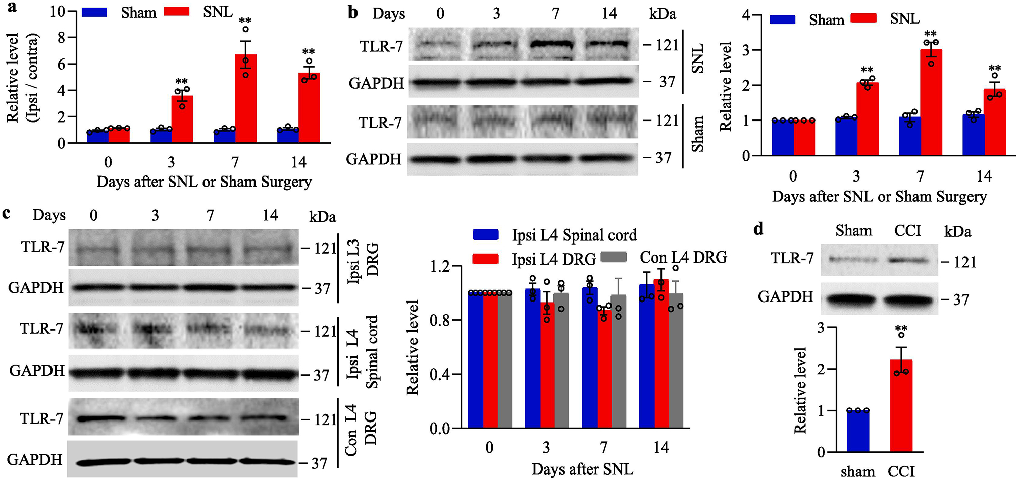
Fig. 1.

Peripheral nerve injury-induced increases in Tlr7 mRNA and TLR7 protein in the injured dorsal root ganglion (DRG). (a) Tlr7 mRNA expression in the ipsilateral L4 DRG of mouse after SNL or sham surgery. n = 3 biological repeats (12 mice)/time point/group. **P < 0.01 vs the corresponding control group (0 day). Two-way ANOVA with repeated measures followed by post hoc Tukey test. (b) Expression of TLR7 protein in the ipsilateral L4 DRG of mouse after SNL or sham surgery. Representative Western blots (left panels) and a summary of densitometric analysis (right graphs) are shown. n = 3 biological repeats (12 mice)/time point/group. **P < 0.01 vs the corresponding control group (0 day). Two-way ANOVA with repeated measures followed by post hoc Tukey test. (c) Expression of TLR7 protein in the ipsilateral L3 (intact) DRG, contralateral L4 DRG and ipsilateral L4 spinal cord of mouse after SNL. Representative Western blots (left panels) and a summary of densitometric analysis (right graphs) are shown. n = 3 biological repeats (12 mice)/time point. (d) Expression of TLR7 protein in the ipsilateral L3/4 DRGs on day 7 after CCI or sham surgery. n = 3 biological repeats (6 mice)/group. **P < 0.01 vs the sham group by two-tailed, independent Student’s t test. Full-length blots are presented in Supplementary Figure 1.
