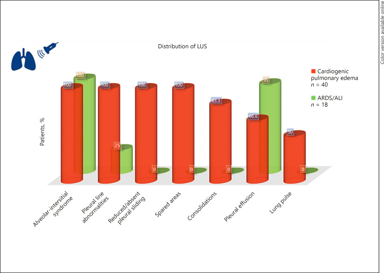
Fig. 2.
LUS patterns in ARDS versus cardiogenic pulmonary edema based on the data from Copetti et al. [18]. Alveolar-interstitial syndrome was defined as the presence of more than 3 B-lines or “white lung” appearance for each examined area. Spared areas were defined as the areas of normal lung pattern in at least one intercostal space surrounded by areas of alveolar-interstitial syndrome. Lung pulse was defined as the absence of lung sliding with the perception of heart activity at the pleural line. LUS, lung ultrasound; ARDS, acute respiratory distress syndrome; ALI, acute lung injury.

