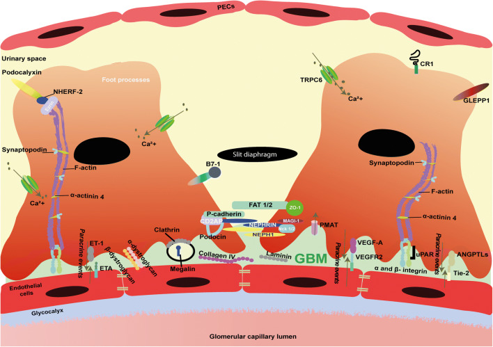
Fig. 1.
Schematic illustration depicting glomerular filtration barrier and molecular overview of podocytes. The three-layered glomerular filtration barrier, consisting of fenestrated endothelial cells, glomerular basement membrane (GBM), and podocytes. Molecules depicted are relevant for the function and characterization of podocytes. These molecules include slit diaphragm, cytoskeleton, and foot processes molecules. Furthermore, hypothesized paracrine signaling molecules (ANGPTLs, VEGF-A, and ET-1) and their receptors, Tie-2, VEGFR2, and ETA, respectively, that are responsible for the crosstalk between endothelial cells and podocytes are shown as well. Finally, podocyte injury markers, such as GLEPP-1, B7-1, CR1, and ezrin are included in the illustration. GBM, glomerular basement membrane; PECs, parietal epithelial cells; ANGPTLs, angiopoietins; CD2AP, CD2-associated protein; MAGI1, membrane-associated guanylate kinase; Nck1/2, non-catalytic region of tyrosine kinase adaptor protein 1/2; NEPH1, same as kin of IRRE-like protein 1 (KIRREL); NPHS1, nephrin; PMAT, plasma membrane monoamine transporter; TRPC6, Transient Receptor Potential Cation Channel Subfamily C Member 6; NHERF-2, Sodium-hydrogen exchange regulatory cofactor NHE-RF2; GLEPP-1, glomerular epithelial protein 1; VEGF-A, vascular endothelial growth factor A; VEGFR2, vascular endothelial growth factor A; ZO-1, zonula occludens-1; α-ACTN4, alpha actinin 4; Ca2+, calcium; ET-1, endothelin 1; ETA, endothelial ET-1 receptor A; Tie-2, tyrosine kinase receptor 2; uPAR, urokinase plasminogen activator receptor

