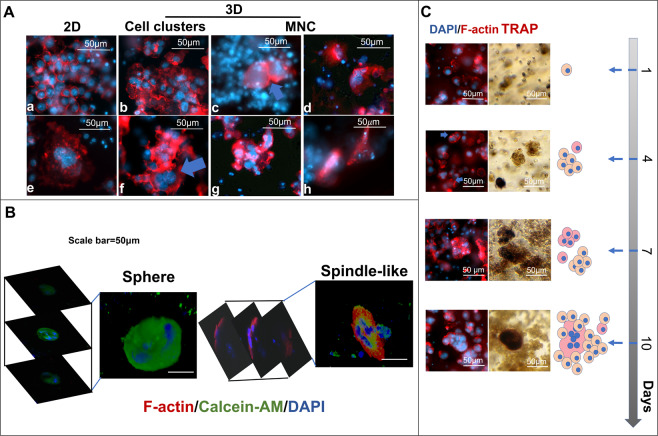
Figure 2.
Morphological alteration of Raw264.7 cell clusters and fused cells by the 3D matrix. (A) The morphology of Raw264.7 cell-derived clusters and fused cells in 2D and 3D cultures (3%: b, f, c, g, and 4.5%: d and h)., F-actin (red, rhodamine phalloidin) and nuclei (blue, DAPI). (B) The 3D constructive images from the z-axis scanning of fluorescence confocal microscope. (C) A series of sequential images obtained from the 3D matrix (3%) embedded Raw264.7 cells cultured for 1,4,7, and 10 days. TRAP(dark red), F-actin (red, rhodamine phalloidin) and nuclei (blue, DAPI).

