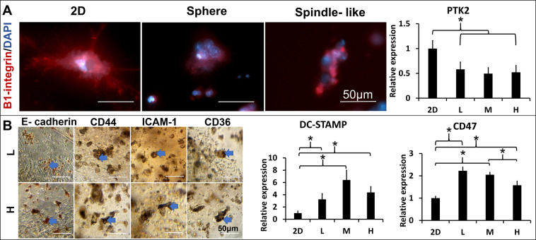
Figure 3.
Localization of the cell-matrix and cell-cell adhesions in the 3D matrices-restrained Raw264.7 cells. (A) The cell-matrix adhesion of Raw264.7 cells derived MNCs in the L or H matrices was identified by the β1-integrin(red) expression with fluorescence immunocytochemistry. (Left); The integrin-mediated focal adhesion gene (PTK2) expression from the 2D and 3D (L, M and H matrices) cultured Raw264.7 cells was quantified by qPCR (n = 3, *p < 0.05) (Right). (B) The cell-cell interactions were identified by stainings of the E-cadherin, CD44, ICAM-1, and CD36 (brown) with peroxidase immunocytochemistry in the L and H matrices. (left). The macrophage fusion competency was quantified by the expression of cells fusion protein genes (DC-STAMP and CD47) from the 2D and 3D (L, M and H matrices) cultured Raw264.7 (right), *p < 0.05.

