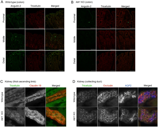
Figure 8.
Immunofluorescence localization of tricellulin in Ildr1 KO and WT mice large intestine and kidney. Representative immunofluorescence images showing angulin-2/ILDR1 (green) and tricellulin (red) antibody staining in each section of the colon for (A) WT mice and (B) Ildr1 KO mice. Scale bar 50 µm. Representative images showing tricellulin in the (C) thick ascending limb (using occludin and claudin 19 as markers) and (D) collecting duct (using occludin and AQP2 as markers) in the kidney of WT and Ildr1 KO mice.

