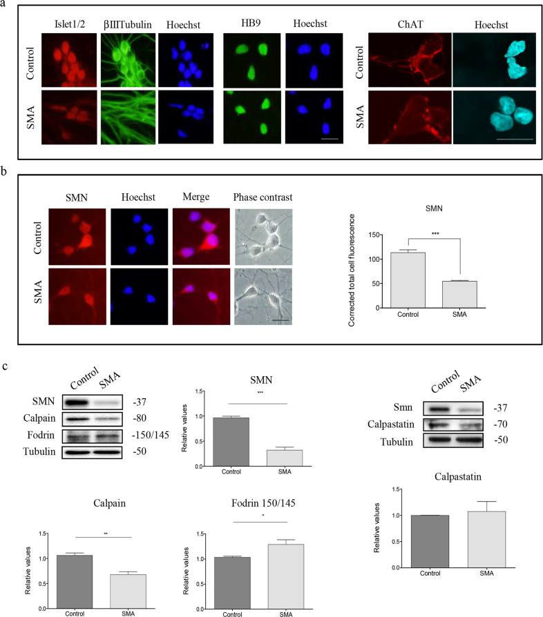
Fig. 3. Calpain protein levels are reduced in human SMA iPSC differentiated MNs.
a Representative immunofluorescence images of 3-day differentiated motoneurons (MNs) in Control and spinal muscular atrophy (SMA), showing Islet 1/2 (red), βIIITubulin (green left section), Hoechst (blue), and HB9 (green right section). b Representative immunofluorescence images of Control and SMA MNs using an anti-SMN antibody and Hoechst staining after 3 days of culture. Graph represents the mean of relative SMN fluorescence (red) measured in cell soma, corresponding to the quantification of three independent experiments ± SEM. Asterisks indicate significant differences using Student t test (p < 0.0001). c Protein extracts of 3-day differentiated human MNs were submitted to western blot analysis and probed with anti-SMN antibody, anti-calpain 1 antibody, anti-fodrin antibody, or anti-calpastatin antibody. Membranes were reprobed with an anti-α-tubulin antibody, used as a loading control. Graphs represent the expression of SMN, calpain, 150/145 fodrin products or calpastatin, corresponding to the quantification of at least three independent experiments ± SEM. Asterisks indicate differences using Student t test (*p < 0.05; **p < 0.001; ***p < 0.0001). Scale bar, 20 μm.

