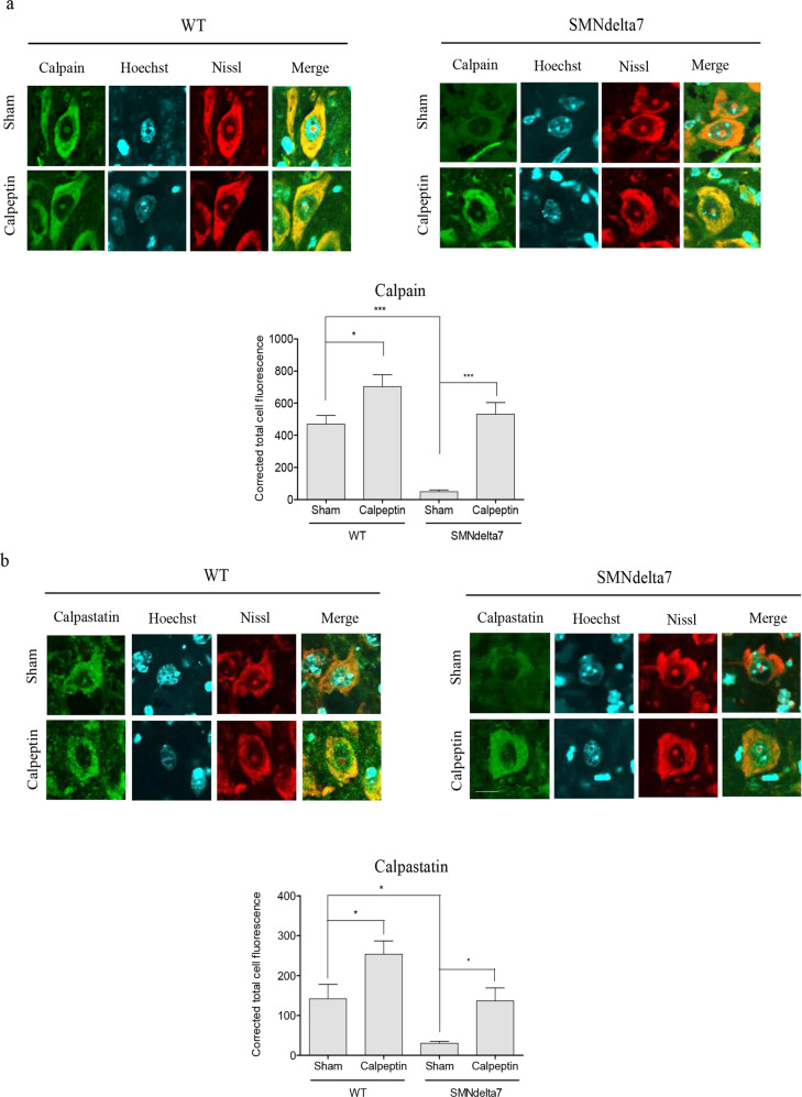
Fig. 5. Calpeptin in vivo treatment increase calpain and calpastatin in SMA spinal cord MNs.
Wild-type (WT) and SMNdelta7 genotyped mice were daily treated with vehicle (Sham) or 6 μg/g of calpeptin (Calpeptin) and sacrificed at 8 post-natal days. Representative immunofluorescence images of ventral horn spinal cord sections of WT and SMNdelta7 vehicle- or calpeptin-treated mice using anti-calpain 1 antibody (green) (a) or anti-calpastatin antibody (green) (b) and Nissl staining (red). Scale bar, 10 μm. Nissl and Hoechst (blue) staining were used to identify MNs soma and nucleus, respectively. Graph represents the mean of relative calpain (a) or calpastatin (b) fluorescence measured in MNs soma, corresponding to the quantification of at least 50 neurons per condition from sections of minimum 3 treated animals ± SEM. Asterisks in a and b indicate significant differences using one-way ANOVA with Bonferroni multiple comparisons post-test (*p < 0.05; **p < 0.001; ***p < 0.0001). Images were acquired with an FV10I confocal microscope (Olympus) using the ×60 objective and the same microscopy settings. Images were not submitted to any post-capture manipulation.

