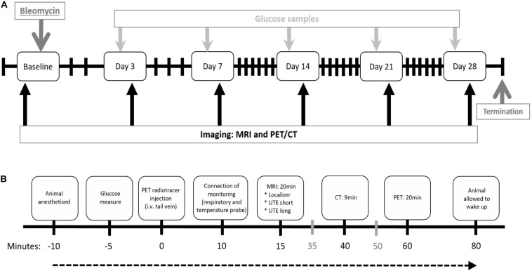
FIGURE 1.
Study layout and imaging workflow chart. (A) Study layout of the DIILD model with bleomycin-induced lung injury in Sprague–Dawley male rats. Imaging was performed at baseline (scan 1) before the intratracheal (i.t.) administration of bleomycin. Control rats received saline as i.t. challenge. Imaging sessions occurred at day 0, 3, 7, 14, 21, and 28 post challenge. Before every scan session, blood glucose was measured. Termination occurred at day 28, after the final imaging session. (B) Workflow for each imaging session. Events expressed in gray (35 and 50 min after injection) indicate when the following rat is prepared while the scan of the previous rat is being performed. i.v. = intravenous.

