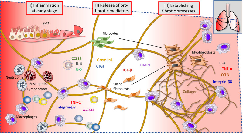
FIGURE 10.
Summary of events during ongoing lung injury, with related cells and markers involved at the injury site of the lung tissue. Immune cells and gene markers that were analyzed throughout this study are presented in a probable scenario during lung injury, at a microscopic viewed site within the lung tissue. The early phase of bleomycin-triggered injury induced immune cell activation and infiltration, observed significantly elevated at day 3–7 post challenge (I). Simultaneously, inflammatory cytokines and pro-fibrotic markers assessed to be significantly increased at this point were several important markers known to be involved in driving the fibrosis progression (II). The assessed lesions by histology, indicated collagen-rich sites and dense fibrotic lesions, also evident by MRI and PET imaging. The later time points (III) indicated a second phase of macrophage infiltration (day 28) in BAL, while several genes were upregulated in particular during the last day of this model (EMT, epithelial–mesenchymal transition).

