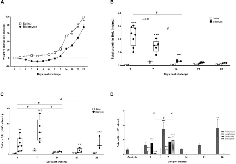
FIGURE 2.
Characterization of the DIILD model, with bleomycin-induced lung injury. (A) Total body weight change in rats over time, expressed as% change compared to their baseline weight. From the BALF analysis total protein concentration and immune cell were assessed at different time points. (B) The total protein reflected plasma exudation and edema at initial inflammatory phase of the bleomycin model (day 3–7 post challenge). (C) The total cell count from BALF was peaking at day 7. Significant increase of cells was also observed at the latest time point (day 28), indicating a second phase of immune cell infiltration. (D) Differential cell counts indicating which cells dominate the various time points. The cells induced at the latest time point (day 28) are only macrophages although present at levels similar to the acute inflammation time point (day 7). Significance is indicated by ** when p < 0.01 and *** when p < 0.001. The comparison of various time points between bleomycin-challenged groups is expressed as # when p < 0.05.

