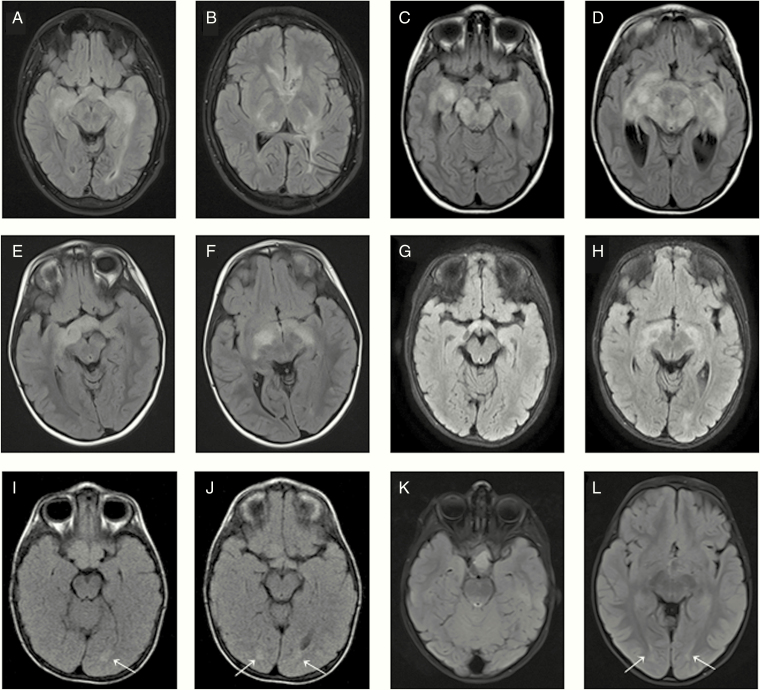
Figure 1.
Axial FLAIR images showing infiltrative OPG spreading at the entire optic pathway corresponding to patients P1 (A, B), P6 (C, D), P7 (E, F), and P9 (G, H). Axial FLAIR images showing OPG mainly localized at chiasm with slight bilateral changes (arrows) at the optic radiation fibers corresponding to patients P14 (I, J) and P15 (K, L).

