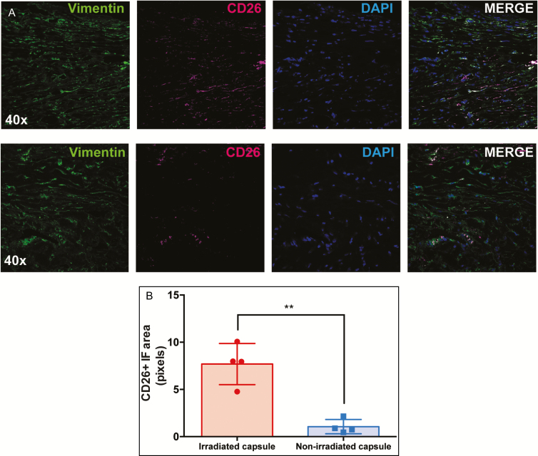
Figure 2.
(A) Immunohistochemical staining of capsule specimens from irradiated (top row) and nonirradiated (bottom row) breast tissue. Cryosections were immunofluorescently stained with vimentin (green) to label fibroblasts and CD26 (magenta) to label the fibrogenic subpopulation of fibroblasts. Nuclei were stained with 4′,6-diamidino-2-phenylindole (blue). Images shown are at 40× magnification. (B) Multiple sections of each specimen were analyzed for co-staining of CD26 and vimentin, which revealed a greater number of CD26-positive fibroblasts in the irradiated capsule (P = 0.0012**).

