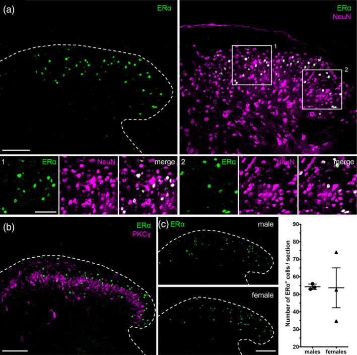
Figure 1.

Estrogen receptor α (ERα) expression in the spinal cord. (a) ERα (green) is expressed by NeuN (magenta) positive neurons in the spinal cord dorsal horn. Insets 1 and 2 depict examples of overlap. (b) ERα is mainly expressed by cells of lamina II of the dorsal horn, with scattered cells both superficially and in deeper laminae (III–V). A subset of PKCγ excitatory interneurons serves as a landmark for inner lamina II. ERα and PKCγ do not overlap. (c) Males and females have comparable numbers of dorsal horn ERα+ cells. Right panel displays quantification from three male and three female mice. Data are presented as mean ± SEM (males: 54 ± 0.88 and females: 54 ± 11). Two‐tailed unpaired t test with Welch's correction for unequal variances: t = 0.05349, df = 2, p = .9622. Dashed lines outline the border of the spinal cord dorsal horn. Scale bar: 100 μm; inset: 50 μm [Color figure can be viewed at wileyonlinelibrary.com]
