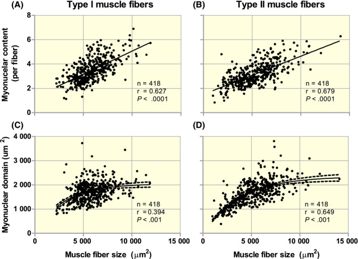
FIGURE 1.

Correlation analysis between type I and type II muscle fibre size and number of myonuclei per fibre (A and B; linear relation) and myonuclear domain size (C and D; logarithmic relation) in percutaneous biopsy samples taken from the vastus lateralis of both healthy adult men (n = 330) and women (n = 88). All samples were collected with subjects at rest following an overnight fast. Muscle fibre size and myonuclear content were determined by immunofluorescent microscopy of muscle cross‐sections. Staining included antibodies for laminin (cell border), MHCI (type I muscle fibres), Dapi (nuclei), Pax7 or NCAM (satellite cells). A Dapi + cell was considered to be a myonucleus when at least 50% of the staining was present within the muscle fibre identified by laminin staining. Muscle satellite cells were identified by Pax7 or NCAM staining and excluded from the myonuclear counts. At least 100 type I and 100 type II muscle fibres per subject were included to make a reliable estimation of myonuclear content
