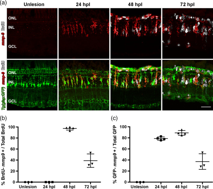
Figure 3.

Following a photolytic lesion, Müller glia and Müller glia‐derived progenitors express mmp‐9. These experiments were performed using the transgenic line Tg[gfap:EGFP]mi2002, in which eGFP is expressed in Müller glia. (a) Triple labeling using in situ hybridization for mmp‐9 (red) and immunostaining for BrdU for dividing cells (white), combined with GFP immunostaining (green). (b) Number of BrdU+ cells that express mmp‐9 in unlesioned animals and lesioned animals at 24, 48, and 72 hpl. (c) Number of GFP+ Müller glia that also express mmp‐9 expression at 24, 48, and 72 hpl. Scale bar equals 25 μm. GCL, ganglion cell layer; INL, inner nuclear layer; ONL, outer nuclear layer [Color figure can be viewed at wileyonlinelibrary.com]
