Figure 10.

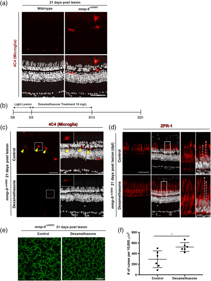
Late anti‐inflammatory treatment rescues defects of regenerated cones in mutants at 21 dpl. (a) Immunostaining for microglia marker, 4C4 in Wild‐type (0.1 ± 0.3 cells per 400 μm, n = 4) and mutant (4.0 ± 2.2 cells per 400 μm, n = 4, p = .0148) at 21 dpl. (b) Experimental paradigm for the photolytic lesions and Dexamethasone treatment. (c) Immunostaining for 4C4 in control (top) and Dex‐treated retinas (bottom). Inserts illustrate ameboid (top) and ramified (bottom) microglia. (d) Immunostaining with ZPR‐1 for red‐green double cones in control (top) and Dex mutants (bottom) 21 dpl. Insets illustrate differences in the lengths of the cone photoreceptors (dashed lines; control, 22.1 ± 7.2 μm, n = 40 cells; Dex‐treated, 29.7 ± 4.8 μm, n = 69 cells; p < .001) (e) Wholemounts of mutant retinas immunostained for ZO‐1. Control retina is left; Dex‐treated retina is right. (f) Number of regenerated cones in control (292.20 ± 158.11 cones; n = 6) and Dex‐treated retinas (522.79 ± 82.55 cones; n = 6) at 21 dpl. Scale bars equal 50 μm in panel (a), 40 μm in panels (c) and (d), and 10 μm in (e) [Color figure can be viewed at wileyonlinelibrary.com]
