Figure 5.

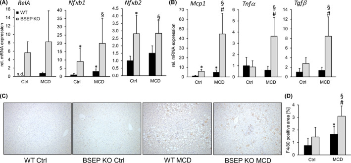
Loss of BSEP aggravates hepatic inflammation under MCD feeding. (A) Expression of NFκB subunits, RelA, Nfκb1 and Nfκb2, was assessed by qPCR. All genes were increased in BSEP KO mice at baseline and tended to be further enhanced by MCD feeding. (B) Inflammatory markers, Mcp1, Tnfα and Tgfβ, were assessed by qPCR. Expression of all genes was markedly increased in MCD‐fed BSEP KO mice. (C) Representative immunohistochemistry for F4/80+ cells of liver specimens of control and MCD‐fed WT and BSEP KO mice (10× magnification) shows increased inflammation in MCD‐fed BSEP KO mice. This observation was confirmed by computational quantification of the sections (D) BSEP KO MCD‐fed mice show the highest levels of F/80 positive area. *Indicates a significant difference in untreated WT controls (Ctrl); § indicates a significant difference in MCD‐fed WT; # indicates a significant difference in BSEP KO Ctrl; P < .05
