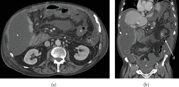
Figure 3.

Abdominal computed tomography after Ladd's procedure in (a) transverse and (b) coronal planes demonstrating intraabdominal loculated ascites (∗) in Morrison's pouch, right and left paracolic gutters, and pelvis. There is resolution of small-bowel obstruction after Ladd's procedure with the cecum (c) positioned in the left upper quadrant medial to the descending colon (d).
