Figure 2.

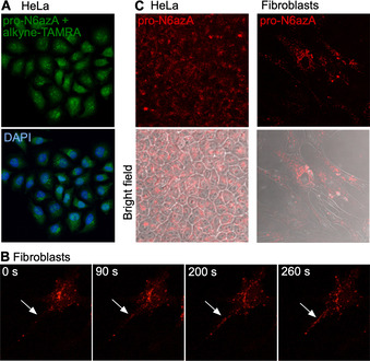
Fluorescence imaging. A) Imaging with pro‐N6azA and alkyne‐TAMRA in fixed HeLa cells. B) Directional transport of labelled proteins towards the processes of fibroblasts imaged in living fibroblast cells by using a time‐lapse camera. White arrows point to the process of the cell. C) Pro‐N6azA probe staining in living HeLa and fibroblast cells with DBCO‐TAMRA 1 d after SPAAC. Control containing HeLa cells not treated with the probe, but treated with DBCO‐TAMRA shows minimal background labelling (Figure S3).
