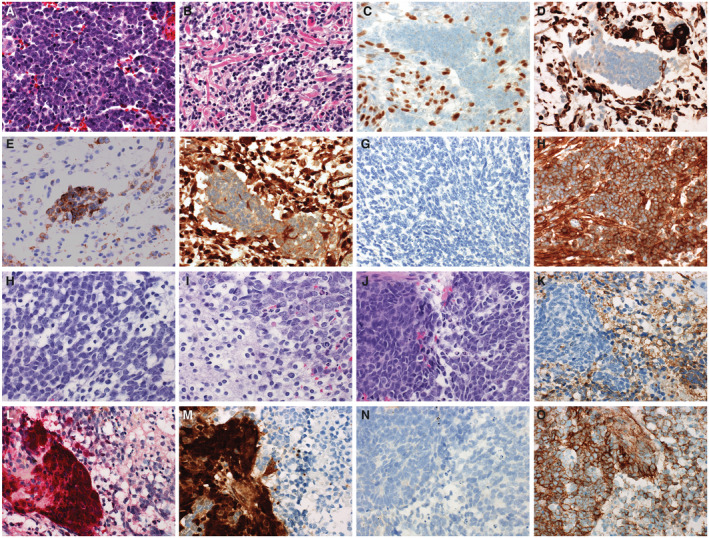
Figure 2.

Medulloblastomas with divergent differentiation most often are characterized by myogenic. Here, a case of medulloblastoma with myogenic differentiation is presented (A‐H). These tumors typically show histology in regions that are typical of other medulloblastomas with primitive elements, often with some anaplastic features (A). In some, overt myogenic differentiation can be observed and can be detected by the presence of “strap” cells (B). Myogenic elements in medulloblastoma with myogenic differentiation can be detected by immunohistochemistry to myogenin (C) or desmin (D). The neuronal elements of medulloblastoma with myogenic differentiation is immunoreactive for synaptophysin (E). The panel of stains used for molecular subgrouping yield a discordant phenotype, showing immunoreactivity for YAP1 (F), no staining for GAB1 (G), and cytoplasmic only staining for beta‐catenin (H). Divergent differentiation can also take the form of melanotic differentiation (I‐P). This case demonstrates the more typical primitive elements of medulloblastoma (I) and neurocytic differentiation (J). A mixed population of the neuronal and melanotic elements can be seen in (K). The neuronal element is immunopositive for synaptophysin (L), whereas the melanotic element is detectable with antibodies to HMB45 (M). Medulloblastomas with divergent melanotic differentiation also yield a discordant pattern on the molecular subgrouping stains, demonstrating immunoreactivity for YAP1 in the melanotic element (N), no significant immunoreactivity for GAB1 (O), and cytoplasmic only staining for beta‐catenin (P).
