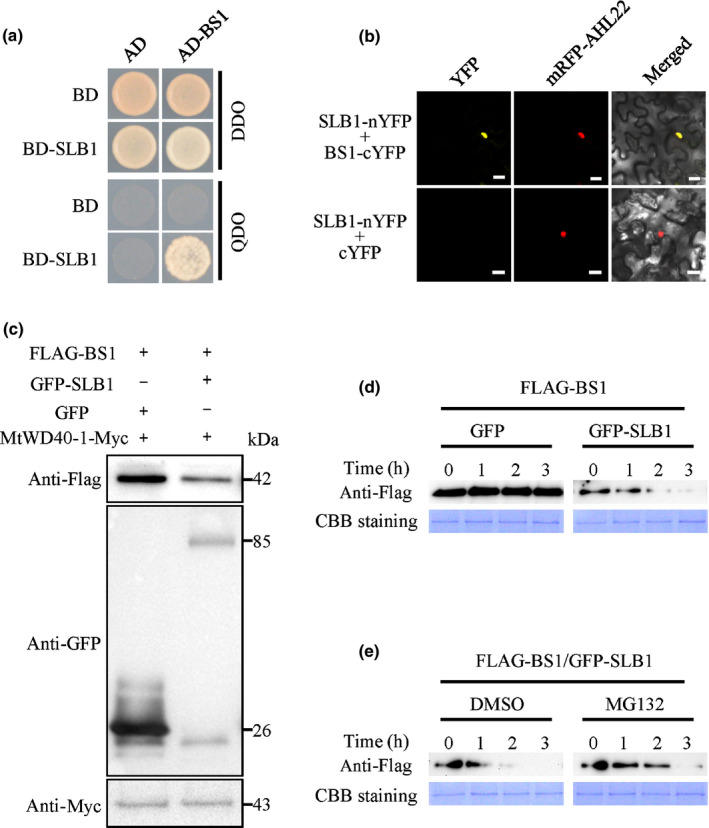
Figure 5.

Medicago truncatula SLB1 interacts with and targets BS1 for degradation. (a) Interaction between SLB1 and BS1 in an Y2H assay. Auxotrophic growth indicates the interaction of each protein. DDO and QDO indicate SD/−Trp/−Leu and SD/−Trp/−Leu/−His/−Ade, respectively. (b) Interaction between SLB1 and BS1 in Nicotiana benthamiana leaf epidermal cells using a BiFC assay. AHL22 was used as a nuclear localisation marker. Bars, 20 μm. (c) SLB1 promotes the degradation of BS1 in vivo. Immunoblotting analysis of total protein corresponding to agro‐infiltrated N. benthamiana leaves with the indicated plasmids. The abundance of FLAG−BS1 was detected using anti‐FLAG antibody, and that of GFP−SLB1 was detected using anti‐GFP antibody. MtWD40‐1‐Myc detection using anti‐Myc antibody served as a loading control. (d) SLB1 promotes the degradation of BS1 in an in vitro protein degradation assay. Protein samples from tobacco leaves coexpressing FLAG–BS1 and GFP–SLB1 or GFP were incubated at 30°C for the indicated times. The abundance of FLAG–BS1 was detected using anti‐FLAG antibody. Coomassie Brilliant Blue (CBB) staining served as a loading control. (e) BS1 degradation is inhibited by MG132. Protein samples from tobacco leaves coexpressing FLAG–BS1 and GFP–SLB1 were treated with MG132 or dimethyl sulfoxide (DMSO, as control) at 30°C for the indicated times. The accumulation of FLAG‐BS1 protein was detected by immunoblotting with anti‐FLAG antibody. CBB staining served as a loading control.
