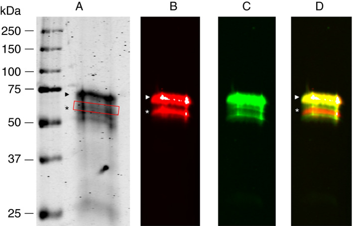
Figure 1.

In‐house purified α2AP visualized by SDS‐PAGE and Western blot. (A) SDS‐gel of purified α2AP. (B) Immunoblot obtained with anti‐Asn‐α2AP. (C) Immunoblot obtained with TC 3AP. (D) Merged anti‐Asn‐α2AP and TC 3AP blots. The relative migration distances of molecular weight marker proteins are indicated on the left. PB‐α2AP is indicated by arrowhead. NPB‐α2AP is indicated by an asterisk, as well as by a red box in (A)
