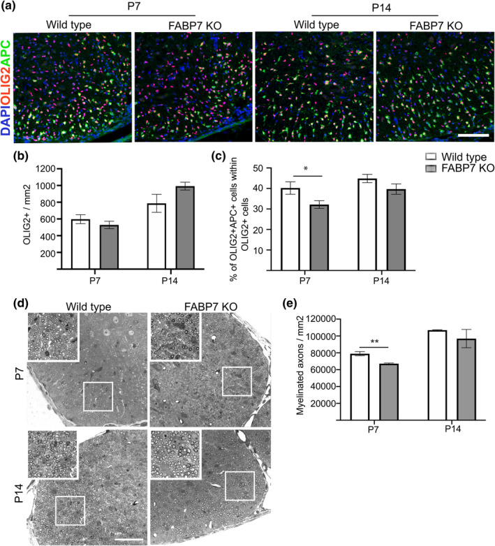
Figure 3.

Developmental myelination in the spinal cord is delayed in Fabp7 knockout mice. (a) Immunohistochemistry staining for OLIG2 and APC (marker of differentiated oligodendrocytes) in the ventral white matter of the spinal cord of WT and Fabp7KO mice at P7 and P14 (Scale bar: 100 μm). (b) Quantification of OLIG2+ cells per area in the whole spinal cord of WT and Fabp7KO mice at P7 and P14 (unpaired Student's t‐test, p = .3430 (P7) and p = .1089 (P14); n = 5–6, mean ± SEM). (c) Quantification of the percentage of OLIG2+APC+ in the white matter of the spinal cord of WT and Fabp7KO mice at P7 and P14 (unpaired Student's t‐test, p = .0435 (P7) and p = .1439(P14); n = 5–6, mean ± SEM). (d) Toluidine blue staining of the ventral spinal cord in WT and Fabp7KO mice at P7 and P14 (Scale bar: 50 μm). White squares highlight areas shown in the higher magnification insets. (e) Quantification of the number of myelinated axons per area in the ventral white matter at P7 and P14 (unpaired Student's t‐test, p = .009 (P7); n = 3 (P7), n = 2 (P14), mean ± SEM)
