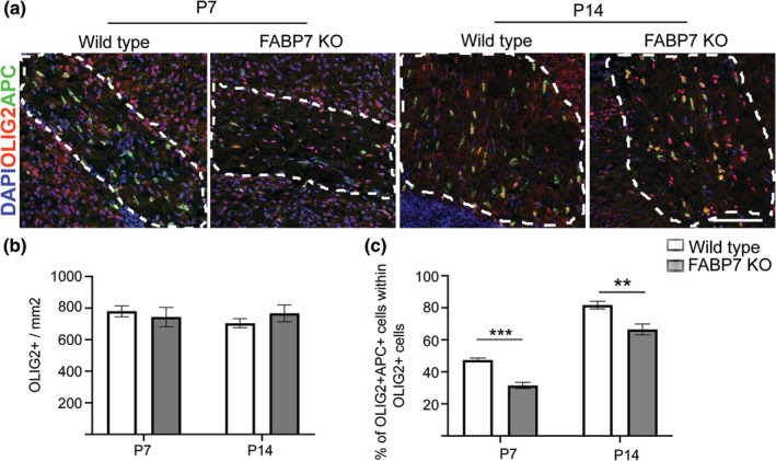
Figure 4.

Developmental myelination in the brain is also delayed in Fabp7 knockout mice. (a) Immunohistochemistry staining for OLIG2 and APC in the corpus callosum of WT and Fabp7KO mice at P7 and P14 (Scale bar: 100 μm). (b) Quantification of OLIG2+ cells per area in the corpus callosum of WT and Fabp7KO mice at P7 and P14 (unpaired Student's t‐test, p = .9307 (P7) and p = .3601 (P14); n = 4–5, mean ± SEM). (c) Quantification of the percentage of OLIG2+APC+ cells in the white matter of the corpus callosum of WT and Fabp7KO mice at P7 and P14 (unpaired Student's t‐test, p = .0001 (P7) and p = .0045 (P14); n = 4–5, mean ± SEM)
