Figure 5.

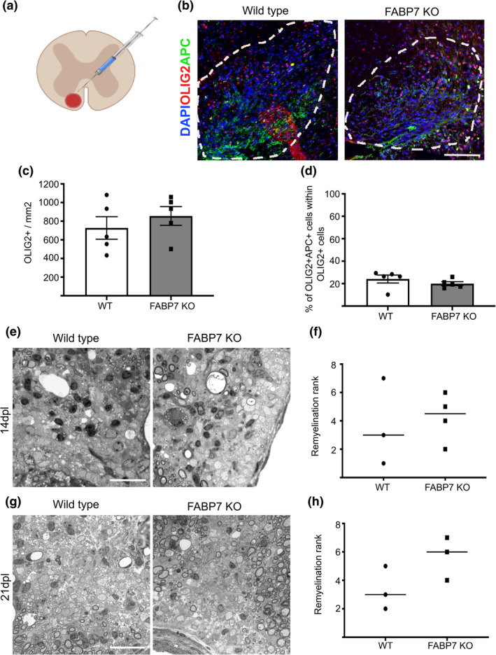
FABP7 is not essential for remyelination. (a) Schematic drawing of lysolecithin induced demyelination in the ventral white matter of the spinal cord. (b) Immunohistochemistry staining for OLIG2 and APC in the lesion in the ventral white matter of WT and Fabp7KO mice at 14 days post lesion (dpl) (Scale bar: 100 μm). (c) Quantification of OLIG2+ cells per area in the lesion of WT and Fabp7KO mice at 14dpl (unpaired Student's t‐test, p = .4371; n = 5, mean ± SEM) (d) Quantification of the percentage of OLIG2+APC+ cells in the lesion of WT and Fabp7KO mice at 14dpl (unpaired Student's t‐test, p = .3154; n = 5, mean ± SEM) (e,g) Toluidine blue staining of the lesion in WT and Fabp7KO mice at 14dpl (e) and 21dpl (g) (Scale bar: 25 μm). (f,h) Ranking analysis of remyelination efficiency in WT and Fabp7KO mice at 14dpl (f) and 21dpl (h) (U‐Mann–Whitney test, p = .8571 (f), p = .2000 (h); n = 3‐4, mean ± SEM)
