
Figure 2.
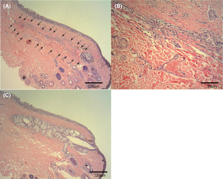
Hematoxylin‐eosin staining of resected eyelid tissue revealed the absence of normal gland lobular and ductal structure at the dropout site (arrows). Original magnification: ×100 (A) or × 400 (B). The histology of tissue corresponding to normal glands on meibography revealed a normal lobular structure of meibomian glands (C)