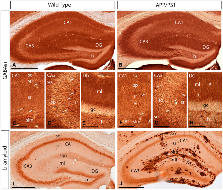
Figure 3.

Regional and cellular distribution of GABAB1 in wild‐type and APP/PS1 mice. A‐H. Immunoreactivity for GABAB1 in the hippocampus of wild‐type and APP/PS1 mice at 12 months of age using a pre‐embedding immunoperoxidase method at the light microscopic level. In the CA1 and CA3 regions and dentate gyrus (DG), GABAB1 immunoreactivity was very similar both in the wild‐type and the APP/PS1 mice, regardless of accumulation of amyloid plaques (asterisks). Labeling for GABAB1 showed the highest intensity in the stratum lacunosum‐moleculare (slm) and molecular layer (ml) and weaker in the strata oriens (so) and radiatum (sr). Immunoreactivity for GABAB1 was also detected in interneurons throughout all layers (white arrows), with similar distribution pattern and labeling intensity in wild‐type and APP/PS1 mice. I,J. Immunoreactivity for β‐amyloid in wild‐type and APP/PS1 mice at 12 months of age, showing high accumulation of Aβ throughout all layers of the hippocampus. Abbreviations: CA1 region of the hippocampus; CA3, CA3 region of the hippocampus; DG, dentate gyrus; so, stratum oriens; sp, stratum pyramidale; sr, stratum radiatum; slm, stratum lacunosum‐moleculare; ml, molecular layer; gc, granule cell layer; h, hilus. Scale bars: A,B,I,J 200 µm; C‐H, 100 µm.
