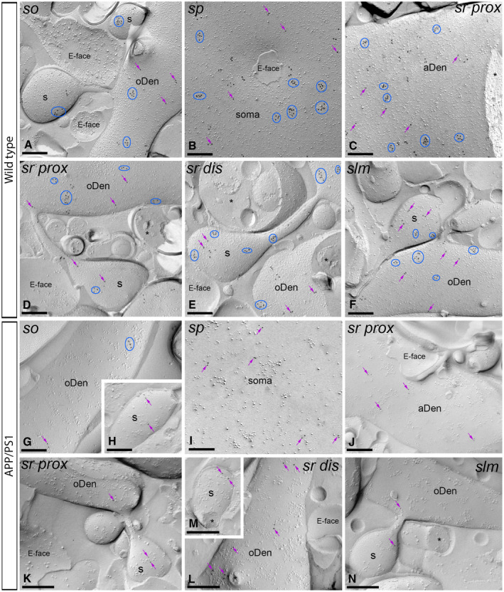
Figure 6.

The density of plasma membrane of GABAB1 is reduced in the hippocampus of APP/PS1 mice at 12 months. Electron micrographs of the CA1 region showing immunoparticles for GABAB1 along the membrane surface of pyramidal cells, as detected using the SDS‐FRL technique. A‐F. In wild type, clusters of GABAB1 immunoparticles (blue ellipses/circles) associated with the P‐face were detected in dendritic shafts, illustrated here for oblique dendrites (oDen) and dendritic spines (s) of pyramidal cells in all strata of the CA1 region. Lower density of immunoparticles for GABAB1 was also detected scattered (purple arrows) outside the clusters. G‐N. In APP/PS1, surface GABAB1 immunoparticles were detected in the P‐face of all compartments of CA1 pyramidal cells. However, GABAB1 immunoparticles were detected at lower frequency forming clusters (blue ellipses/circles), being showing a scattered distribution (purple arrows). Fractured spine necks and other cross‐fractures are indicated with asterisks (*). The E‐face is free of any immunolabeling. Scale bars: A‐N, 0.2 μm.
