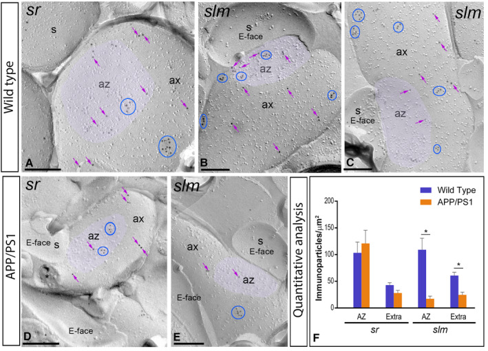
Figure 8.

Presynaptic localization of GABAB1 in the hippocampus in wild‐type and APP/PS1 mice. Electron micrographs showing immunoparticles for GABAB1 in presynaptic compartments in the strata radiatum (sr) and lacunosum‐moleculare (slm) of the CA1 region of the hippocampus at 12 months of age, as detected using the SDS‐FRL technique. A‐C. In wild type, immunoparticles for GABAB1 were found within the active zone (az, purple overlay), recognized by the concave shape of the P‐face and the accumulation of IMPs, and along the extrasynaptic site of axon terminals (ax), forming clusters (blue ellipses/circles) and also detected scattered (purple arrows) outside the clusters. D,E. In APP/PS1, fewer immunoparticles for GABAB1, forming clusters (blue ellipses/circles) or scattered (purple arrows), were detected within the active zone (az, purple overlay) and along the extrasynaptic plasma membrane of axon terminals (ax). Cross‐fractures are indicated with asterisks (*). F. Densities of immunoparticles for GABAB1 in presynaptic compartments in the sr and slm in wild‐type and APP/PS1 mice. No differences were detected in densities of GABAB1 immunoparticles in the sr (WT: AZ = 103.31 ± 20.16 immunoparticles/µm2 and Extra = 42.97 ± 4.47 immunoparticles/µm2; APP: AZ = 120.95 ± 24.58 immunoparticles/µm2; Extra = 28.08 ± 4.96 immunoparticles/µm2), but significant differences were detected in the slm (WT: AZ = 109.03 ± 21.62 immunoparticles/µm2 and Extra = 60.79 ± 6.11 immunoparticles/µm2; APP: AZ = 17.32 ± 4.99 immunoparticles/µm2; Extra = 24.64 ± 5.17 immunoparticles/µm2) (Two‐way ANOVA test and Bonferroni post hoc test, *P < 0.05). Error bars indicate SEM. Scale bars: A‐E, 0.2 μm.
