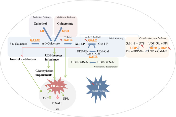
Figure 1.

Involved pathways, pathophysiological agents and targets for treatment in hereditary galactosemia, studied in cellular and animal models , indicating a treatment approach that has been evaluated in the cellular and animal models.
  , inhibition.
  , induction. GALK deficiency was studied in yeast (Y), fruitfly (F) and mouse (M). GALT deficiency was studied in patient cell cultures (C), bacteria (B), yeast, fruitfly, zebrafish (ZF) and mouse. GALE deficiency was studied in bacteria, yeast, nematode (N) and fruitfly. AR, aldose reductase, ER, endoplasmic reticulum, Gal‐1‐P, galactose‐1‐phosphate, GALE, UDP‐galactose 4′‐epimerase, GALK, galactokinase, GALT, galactose‐1‐phosphate uridylyltransferase, GALM, galactose mutarotase, GDH, galactose dehydrogenase, Glc‐1‐P, glucose‐1‐phosphate, PI3/Akt, phosphatidylinositol‐4,5‐bisphosphate 3‐kinase/protein kinase B, PPi, inorganic pyrophosphate, ROS, reactive oxygen species, RP, ribosomal protein, UDP‐Gal, uridine diphosphate‐galactose, UDP‐GalNAc, UDP‐N‐acetylgalactosamine, UDP‐Glc, uridine diphosphate‐glucose, UDP‐GlcNAc, UDP‐N‐acetylglucosamine, UGP, UDP‐glucose pyrophosphorylase, UPR, unfolded protein response, UTP, uridine‐5′‐triphosphate
