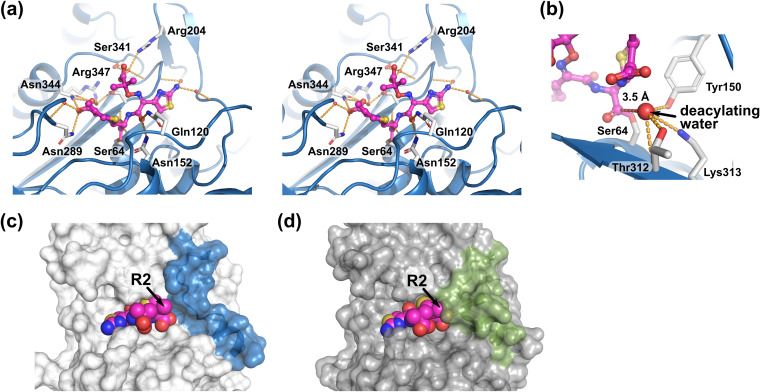
FIG 4.
Ceftazidime recognition by AmpCEnt385. (a) Stereo view of the ceftazidime binding. The ceftazidime molecule as shown in ball-and-stick representations colored magenta. Hydrogen bonds are shown as orange dashed lines. Residues and the water molecules participating the hydrogen bond network are shown as white stick and red spheres, respectively. (b) The deacylating water molecule is shown as a large red sphere. Red dashed line indicates the distance between C-8 atom of ceftazidime and the deacylating water molecule. (c and d) Comparison of the substrate-binding sites of the AmpCEnt385-CAZ complex and the AmpCEc-CAZ complex. Transparent molecular surfaces of AmpCEnt385 (c) and AmpCEc (d) are colored white and gray, respectively. To clarify the difference, the colors of the V283–V294 residue structures, including R2 loop in AmpCEnt385, and corresponding residues in AmpCP99 appear in blue and green, respectively. Arrowheads indicate the R2 side chain positions of ceftazidime.

