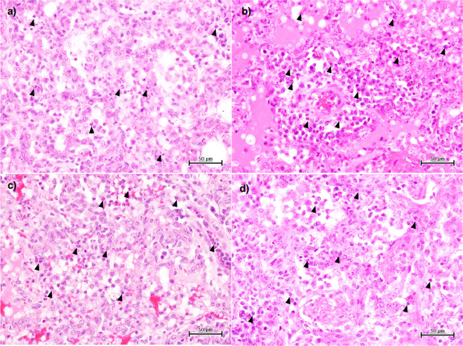
FIG 2.
Viral pneumonia in cynomolgus macaques challenged with A/black swan/Akita/1/2016 (H5N6). Shown are images of H&E staining of lung tissues collected 7 days after virus infection. Representative photos of cynomolgus macaques treated with saline (a), oseltamivir (b), peramivir (c), and amantadine (d) are shown. Black arrowheads point to neutrophils.

