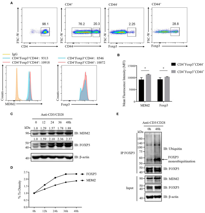
Figure 7.
TCR/CD28 signaling enhances MDM2 expression and MDM2-mediated ubiquitination of FOXP3 in human Treg cells. (A) MDM2 and Foxp3 expression levels were examined in activated Treg cells (CD4+Foxp3+CD44high) and non-activated Treg cells (CD4+Foxp3+CD44low) of CD4+ T cells isolated from lymph nodes of WT C57BL/6 mice. (B) MDM2 and Foxp3 expression levels were analyzed as demonstrated in (A). (C,D) In vitro differentiated human Treg cells were rested (which meant Treg cells were removed from anti-CD3/CD28 beads, with addition of 100 U/ml IL-2) for 2 days, and were re-stimulated with anti-CD3 (1 μg/ml) and anti-CD28 (1 μg/ml) antibodies in the presence of 100 U/ml IL-2 for 0, 12, 24, 36, or 48 h. The endogenous MDM2 and FOXP3 expression levels were detected by Western blot assay. (E) Human Treg cells were rested for 2 days and were stimulated with anti-CD3 (1 μg/ml) and anti-CD28 (1 μg/ml) antibodies in the presence of 100 U/ml IL-2 for 0 or 48 h, followed by the assessment of FOXP3 ubiquitination and FOXP3 and MDM2 expression, through utilizing immunoprecipitation assay and Western blot assay. The arrow indicates FOXP3 monoubiquitination. The above data are derived from more than three independent experiments. *p < 0.05.

