Figure 3.

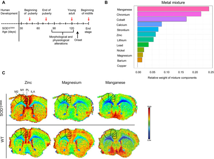
Metal dyshomeostasis in a mouse model of ALS. (A) Lower mandibles and whole brains were harvested from SOD1G93A and littermate control mice at 47, 71, 120, and ~160 days of age (red arrows). Corresponding developmental periods in humans are indicated (top). Morphological and physiological alterations such as neuromuscular junction dysfunction and motor skill deterioration in SOD1G93A mice develop before disease onset. (B) LA‐ICP‐MS was used to determine metal concentrations in teeth and mixture analyses were performed by WQS to identify the relative weights (importance) of each metal in the mixture when comparing transgenic to wild‐type (WT) mice by groups (P = 0.037; SOD1G93A n = 14; WT n = 15). (C) Brain distribution of biometals is altered in transgenic mice. Representative metal spatial concentration for zinc, magnesium, and manganese by LA‐ICP‐MS on 30 µm anterior sections of coronal mouse brain from SODG93A and WT animals (end‐of‐stage). Anatomical regions of interest are delineated. M1, primary motor cortex; M2, secondary motor cortex; PL, prelimbic area; ILA, infralimbic area. For elemental maps, pixel size is approximately 30 × 30 µm.
