Figure 1.

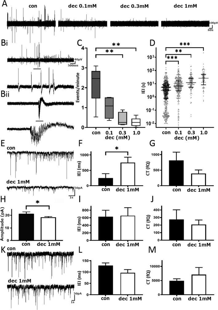
Decanoic acid reduces spontaneous epileptiform activity in pediatric epilepsy tissue samples. (A) Example trace of local field potential recording before and after consecutive addition of DEC. Scale bar 100 µV versus 60 sec. (B) Two examples of spontaneous epileptiform activity from separate pediatric human tissue recordings. Black bar denotes zoomed in section of events. Scale bars (top) 40 µV versus 500 msec, (bottom) 100 µV versus 2 sec. (C) Pooled events per minute under various conditions. (D) The interevent interval in different concentrations of DEC. (E) Example trace of sEPSCs from pediatric human tissue before (top) and after (bottom) treatment with 1 mmol/L DEC. Scale bar 50 pA versus 1 sec. (F) Pooled mean‐median inter‐event interval in 1 mmol/L DEC. (G) Pooled sEPSC charge transfer in 1 mmol/L DEC. (H) Pooled mean‐medium amplitude of sEPSCs in 1 mmol/L DEC. (I) Pooled inter‐event interval of mEPSCs with 1 mmol/L DEC. (J) Pooled charge transfer of mEPSCs in 1 mmol/L DEC. (K) Example traces of mIPSC recordings pre and post addition of 1 mmol/L DEC. Scale bar 50 pA versus 1 sec. (L) Pooled interevent interval of mIPSCs in control and 1 mmol/L DEC. (M) Pooled charge transfer of mIPSCS after addition of 1 mmol/L DEC. DEC, decanoic acid. *P ≤ 0.05; **P ≤ 0.01; ***P ≤ 0.001.
